Technology
L-PRF™
L-PRF™ is a 3-D autogenous combination of Platelet Rich Fibrin derived from the patients’ blood. In simple terms, it is a biological bandage created from patient’s own blood.
A simplified chair side procedure that creates a thin, compressed layer of platelet rich fibrin that is strong, pliable and suitable for surgical manipulation and suturing. This natural fibrin network is rich in platelets, growth factors and cytokines that are derived from the blood platelets and leukocytes. The presence of these proteins has been reported to produce rapid healing, especially during the critical two week period after placement. This network promotes more efficient cell migration and proliferation without chemical or bovine thrombin additives.
It is indicated but not limited to extraction sockets, sinus and dental ridge augmentation procedures, palatal defects, and maxillary bone atrophy.
Cone-Beam CT
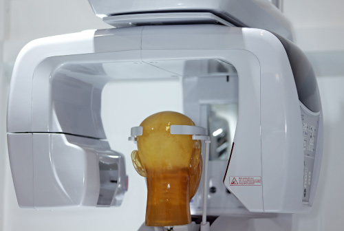
Dr. Patel and his team at Aiken Family Dentistry uses dental cone beam computed tomography (CT), which is a special type of x-ray machine used in situations where regular dental or facial x-rays are not sufficient. This type of CT scanner uses a special type of technology to generate three dimensional (3-D) images of dental structures, soft tissues, nerve paths and bone in the craniofacial region in a single scan. Images obtained with cone beam CT allow for more precise treatment planning.
With cone beam CT, an x-ray beam in the shape of a cone is moved around the patient to produce a large number of images, also called views. CT scans and cone beam CT both produce high-quality images.
Cone beam CT provides detailed images of the bone and is performed to evaluate diseases of the jaw, dentition, bony structures of the face, nasal cavity and sinuses.
Laser Dentistry
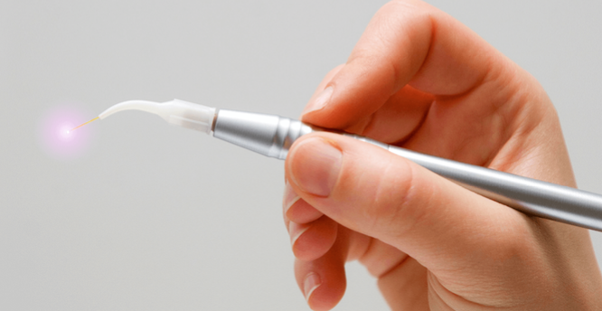
Dental Diode lasers often used in dentistry for procedures that involve cutting or contouring oral soft and hard tissues. At Aiken Family Dentistry, Dr. Patel uses soft-tissue laser as a clinical tool, to perform a wide range of clinical treatment procedures such as troughing around preparations, crown lengthening, curettage of gingival sulcus and treating periodontal disease, sterilizing endodontic canals, fibroma removal, teeth whitening, frenectomy and peri-implantitis treatment; as it is capable of creating precision cuts in gingiva and other soft tissues while also eliminating bleeding at the site and reducing the healing time for the patient.
Intraoral Cameras
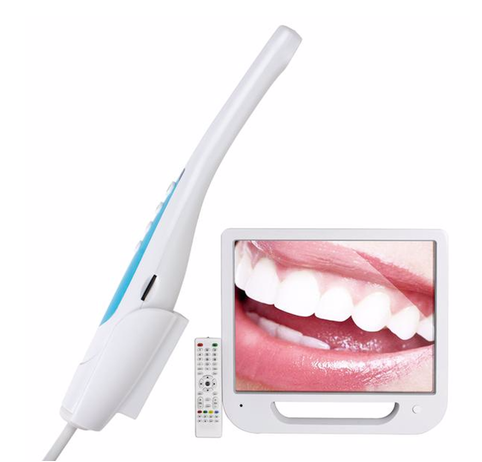
An intraoral camera allows Dr. Patel and his team at Aiken Family Dentistry to capture high quality diagnostic color images of oral cavity allowing for easier and more accurate diagnosis of potential oral health problems.
These crystal clear images are then transmitted onto a patient monitor for communicating, educating, and explaining their diagnoses during consultations.
Using special software, Dr. Patel and his team are able to enlarge these images, freeze them, or view them in real time.
It also allows us at Aiken Family Dentistry to create accurate documentation of a patient’s dental condition and obtain a quicker response from his or her insurer, who will have clear visual evidence to confirm the results of our diagnostic tests.
Digital X-Rays
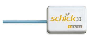
Dr. Patel and his team at Aiken Family Dentistry uses digital radiology in diagnostic exams as their commitment in providing patients with the safest and most effective way of having dental x-xrays.
Digital x-rays are safer, faster, more accurate, and far more biologically conscientious than their traditional counterparts. The digital x-rays require no film, therefore reducing exposure to radiation by 80 percent and eliminating hazardous waste.
Digital x-rays provide high-quality images of your teeth, gums and underlying bone; hence, better diagnosis and outcome.
Patients simply close their mouth on a digital sensor while our computer processes the image within seconds.
Cavitron and Air Polisher
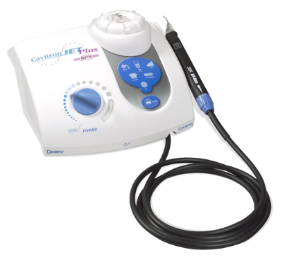
A device that uses high frequency sound waves to vibrate tartar and debris off your teeth. Water then flushes away the bacteria and plaque that build up on your teeth. Air Polisher is a minimally invasive technique which uses a mixture of compressed air, water and fine powder to remove unsightly staining and harmful plaque from the teeth.
Rotary Endodontics
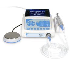
Provides much more comfort to patients during the root canal treatment; more reliable results and faster treatment.
Dental Loupes and LED Light
Enhance or magnify an image to insure the highest quality results are obtained in dental procedures.
Digital Forms
Provides patients with feasibility filling out forms from their personal device.
2-Way Texting
Patients can communicate with our office staff via texting.

